

Accessory navicular
An accessory navicular is an extra bonean “extra” bone compared to normal so not everyone has one. in the foot. Most of the time, it goes unnoticed and does not need to be treated. In other cases, it can cause pain which requires treatment. This pain is often wrongly attributed to an ankle sprain as it is felt near the ankle.
Anatomy and mechanism of injury
In people who do not have an accessory navicular, the tibialis posterior tendonAttaches muscle to bone. inserts into the navicular bone. This bone is relatively large and strong so that it can tolerate the strain on the tendon.
In people who do have an accessory navicular, there are some anatomical differences. The accessory navicular is attached to the navicular bone by a thin, fragile articulation. The tibialis posterior tendon attaches to both of these bones, covering the accessory navicular. As a result of the repeated strain on the tendon, the articulation weakens, mobilises and becomes irritated and inflamed. This inflammation can be seen on an MRI scan and explains the symptoms.
Non-weight-bearingFoot on the ground but not taking the weight of the body. X-rays of the foot can confirm the diagnosis as they are able to show the presence of the extra bone. In atypical cases or when the diagnosis is unclear, an MRI scan can show the inflammation between the navicular bone and accessory navicular, and therefore confirm that this is what is causing the pain.


Symptoms
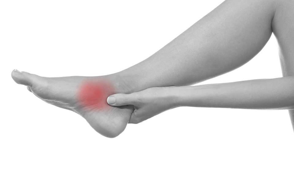
Possible symptoms include: 
- Pain on the inner side of the foot slightly below and in front of the ankle.
- A hard prominence…or lump in common language. in the same area corresponding to the accessory navicular.
- OedemaSwelling caused by a build-up of fluid in the body’s tissues. may also occur.
The pain can worsen on impact (e.g. when the foot hits the ground when running) or if the ankle gets twisted and can therefore end up being wrongly attributed to a foot or medial ankle sprain.
Treatment
Rest, icing and pain medication can all help to treat acute episodes of pain (e.g. if you twist your ankle) but do not prevent it from coming back.
For this reason, surgery is the treatment of choice. The procedure requires a 5 cm incision to be made and takes 45 minutes. It consists of removing the accessory navicular, repairing the tibialis posterior tendon and then reinserting it into the navicular bone using an anchorHarpoon-shaped device that the surgeon fixes in the bone, with threads that come out of the anchor being used to stitch the tendon to the bone.. The incision is then closed using subcuticular continuous sutures which provide the best cosmetic results.
FAQs
- Is rest enough? Rest will relieve the pain initially. However, on resuming your activities, the same process will probably cause the pain to gradually come back.
- An accessory navicular was not found on my X-ray, but it certainly seems like there is one. Make sure that you get X-rays of the foot and not the ankle. They also need to be non-weight-bearingFoot on the ground but not taking the weight of the body. X-rays and NOT weight-bearingFoot on the ground taking the weight of the body. X-rays.
- Are MRI scans routine? No, they are only performed if there is doubt over whether the accessory navicular is causing the pain.
- Can the operation be done as an outpatient procedure? Yes, provided that you have friends or family who can help you.
- Can children have this operation? Yes, provided that the ossification of the navicular bone is at an advanced stage (this usually happens between the ages of 10 and 13). An X-ray can be performed to check for this.
- Is the operation painful? The majority of patients who undergo this surgery do not experience a significant amount of pain. In any case, pain medication will be provided.
- Will I be able to walk after the operation? Yes, but you will have to use crutches for 10 to 15 days.
- When can I play sports again after the operation? After 12 weeks.